Join our lab — Now recruiting graduate students and postdoctoral researchers
We welcome inquiries from prospective graduate students (M.S. and Ph.D. programs) and postdoctoral researchers. Please feel free to contact us. Lab visits are available year-round.
Financial support for graduate students: SPRING (Support for Pioneering Research Initiated by the Next Generation)
Jichi Medical University has been selected for JST’s SPRING program starting in FY2025. The program provides selected Ph.D. students with a living stipend and research funds, allowing them to focus on their studies without financial burden. In addition, students receive multifaceted support, including career-development training and opportunities to strengthen international engagement.
For more information, please visit the Jichi Medical University SPRING program page.
Below are our current major research themes.
Mechanisms of demyelinating disorders and myelin regeneration
Developing an original mouse model of focal demyelination
To precisely evaluate how demyelination impairs motor function, it is important to model demyelination in brain regions directly involved in movement. We developed a mouse model in which focal demyelination is induced in the internal capsule, a key pathway conveying motor commands from the cortex to the spinal cord. This model produces robust motor deficits that recover as remyelination proceeds. It also provides a practical platform for testing compounds that enhance myelin repair.
Yamazaki & Ohno, Acta Histochem Cytochem 57(1), 2024 / Yamazaki et al., J Neurochem 2020 / Yamazaki, Osanai et al., Neurochem Int 164, 2023
Type I collagen as a barrier to remyelination in white matter lesions
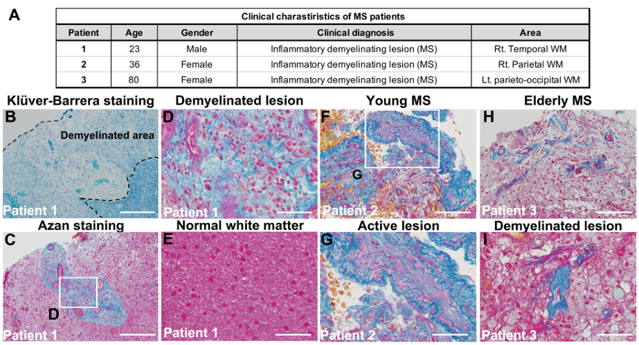
Why do some demyelinated lesions fail to repair efficiently? We found that type I collagen accumulates in demyelinated areas and suppresses the differentiation of oligodendrocytes (the myelin-forming cells), thereby limiting remyelination. We further identified macrophages as a major cellular source of this collagen. Importantly, similar collagen accumulation is observed in brain tissue from patients with multiple sclerosis, highlighting type I collagen as a potential therapeutic target for improving myelin regeneration.
Yamazaki, Azuma, Osanai et al., Cell Death Dis 16(1), 2025 / Inagaki, Fujiwara, Shinohara et al., Histochem Cell Biol 155(4), 2021 / Yamazaki & Ohno, Anat Sci Int 2025
Identifying drugs that promote remyelination
We also pursue drug repositioning to uncover existing compounds that enhance myelin repair. The antihistamine clemastine is known to promote oligodendrocyte differentiation and remyelination, and we are investigating its mechanisms of action and translational potential.
Yamazaki & Ohno, Front Cell Neurosci 19, 2025 / Yamazaki, Osanai et al., Neurochem Int 164, 2023
Experience- and activity-dependent plasticity of myelin
Sensory experience reshapes myelin architecture
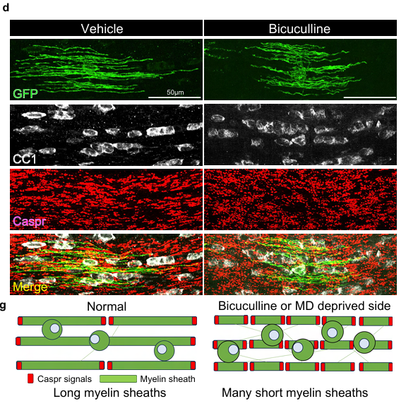
To study how postnatal visual experience influences myelination, we use a dark-rearing (visual deprivation) model. We found that mice reared in the dark during the critical period show structural changes in myelinated axons along the visual pathway. Ongoing work using transcriptomics and related approaches is revealing a role for the neurotransmitter GABA in these experience-dependent myelin changes.
Osanai, Battulga et al., Neurochem Res 47(9), 2022 / Osanai, Battulga, Yamazaki et al., bioRxiv 2025 / Osanai, Shimizu et al., Glia 66(11), 2018
How a single oligodendrocyte myelinates diverse axons
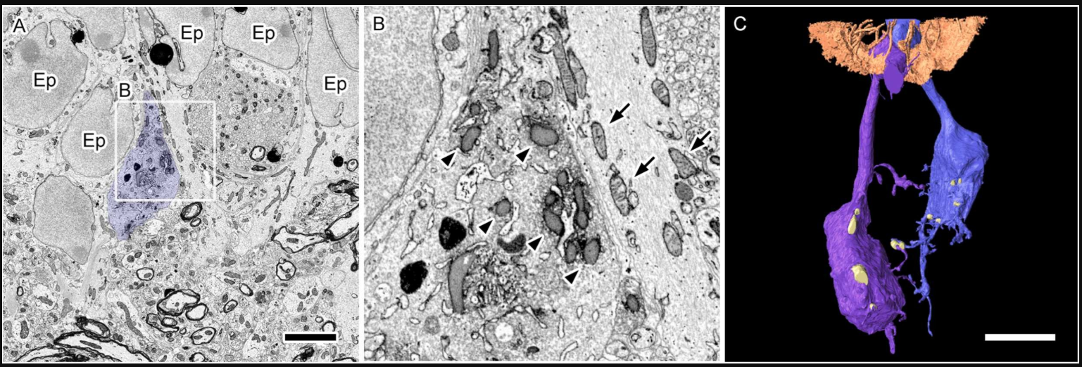
Each oligodendrocyte can myelinate dozens of axons, yet the resulting myelin segments vary widely in length and thickness. Using 3D reconstructions from SBF-SEM datasets, we showed that single oligodendrocytes can simultaneously myelinate axons originating from different brain regions, and that myelin morphology is tuned to axon identity. We view this oligodendrocyte “selectivity” as a key cellular basis for myelin plasticity.
Battulga, Osanai et al., Glia 73(4), 2025 / Osanai, Shimizu et al., Glia 65(1), 2017 / Osanai, Yamazaki et al., Front Cell Dev Biol 10, 2022
Age-associated decline in myelination capacity
Through quantitative analyses of the corpus callosum in aged mice, we found that the production of new oligodendrocytes declines with age, resulting in reduced myelination capacity. We also observe morphological changes in these oligodendrocytes, suggesting links to cognitive aging and neuronal vulnerability.
Looprasertkul, Yamazaki, Osanai & Ohno, Glia 73(11), 2025
Mitochondrial and organelle dynamics
Mitochondrial remodeling in demyelinated axons
What happens to mitochondria when axons lose myelin? Using 3D ultrastructural analysis by SBF-SEM, we found that synaptic terminals in the cerebellum in a chronic demyelination model contain fewer mitochondria, but with increased mitochondrial size, leading to accumulation of swollen mitochondria. We also observed increased contacts between mitochondria and the endoplasmic reticulum (MAM: mitochondria-associated ER membranes) in demyelinated axons, suggesting that altered mitochondria–ER coupling may affect energy metabolism and calcium handling.
Nguyen, Sui et al., Med Mol Morphol 51(4), 2018 / Thai, Nguyen et al., Med Mol Morphol 52(3), 2019 / Sui et al., Adv Exp Med Biol 1190, 2019
Microglial mitochondrial dynamics and neuroinflammation
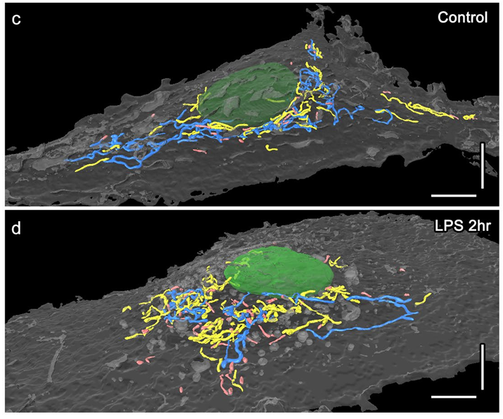
We used SBF-SEM to reconstruct microglial mitochondria in 3D and quantify their morphology. Mitochondrial fission–fusion balance differs substantially across individual microglia, and we showed that these differences are associated with distinct neuroinflammatory phenotypes. We also identified morphological distinctions between monocyte-derived and microglia-derived macrophages, providing an ultrastructural perspective on inflammatory heterogeneity.
Katoh, Wu et al., Sci Rep 7(1), 2017
Brain–periphery metabolic crosstalk underlying cognition and emotion
Chronic stress, immunometabolism, and behavioral outcomes
Stress induces metabolic and immune changes in both the brain and peripheral tissues, and these alterations can be accompanied by emotional dysregulation and cognitive impairment. We showed that chronic stress reduces endogenous pro-resolving lipid mediators that normally help terminate inflammation. We are currently focusing on stress-induced changes in the prefrontal cortex, a key region for executive function and systemic metabolic control.
Higashida, Nagai et al., Sci Rep 2018 / Akiyama, Nagai et al., Sci Rep 2022 / Horikawa, Nagai et al. J Pharmacol Sci 2024 / Nagai, Microscopy 2024
Synaptic mitochondria and inter-individual variability in cognitive aging
Not all individuals age in the same way. We found that oxidative stress in synaptic mitochondria in the prefrontal cortex of aged mice is a key factor underlying individual differences in cognitive decline. Building on our expertise in volume electron microscopy, we are linking synapse-level structural alterations to behavioral outcomes.
Yamada, Nagai et al., bioRxiv 2026, Yamada, Nagai et al., bioRxiv 2026, Yamada, Nagai et al., J Pharmacol Sci 2025
Synapse ultrastructure and morphological homeostasis
Synapses change in number and shape in response to everyday experiences and sleep, yet many aspects of this remodeling remain poorly understood. Using volume electron microscopy, we provided evidence consistent with the idea that sleep downscales synaptic structures in mice, creating capacity for new learning. We also found that prolonged sleep deprivation during early life can irreversibly induce dendritic morphological abnormalities. These findings open avenues for extending volume EM approaches to diverse physiological and pathological contexts.
Nagai, de Vivo et al., Sleep 2017 / de Vivo, Nagai et al., Sleep 2019 / Nagai, de Vivo et al., eNeuro 2021
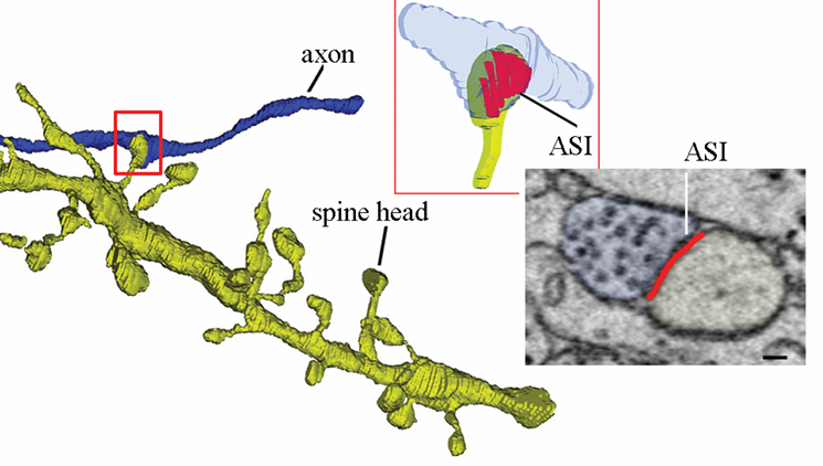
Advancing volume electron microscopy and expanding its applications
Improving imaging quality with conductive resin embedding
In SBF-SEM, charging of the specimen surface can severely degrade image quality. We developed a conductive-resin embedding approach that substantially reduces charging and improves image resolution, enabling the acquisition of high-quality, large-volume 3D datasets.
Nguyen, Thai et al., Front Neural Circuits 12, 2018 / Nguyen, Thai et al., Sci Rep 6, 2016
Three-dimensional circuit reconstruction at ultrastructural resolution
SBF-SEM makes it possible to reconstruct and analyze neural circuits labeled by specific genes or molecular markers at ultrastructural resolution. We also combine this with correlative light and electron microscopy (CLEM)—labeling cells by light microscopy and then examining their ultrastructure by electron microscopy—to achieve a more precise understanding of circuit organization.
Ohno, Karube & Fujiyama, Neurosci Res 2024 / Abe & Ohno, Microscopy 2024 / Ohno et al., Microscopy 65(2), 2016
Collaborations across disciplines
Our volume EM platform supports a wide range of collaborative projects beyond neuroscience, including renal pathology, botany, gastroenterology, dermatology, cardiovascular surgery, and developmental biology. We work with partners to address questions that become accessible only through large-scale 3D ultrastructural datasets—for example, ciliary morphology in polycystic kidney disease models, evo-devo studies of head mesoderm in lampreys, and nuclear migration in tobacco pollen mother cells.
Kumamoto, Kagami et al., Sci Rep 2026 / Onai, Adachi et al., iScience 26(12), 2023 / Mursalimov et al., Front Plant Sci 2021 / Ezure et al., Sci Rep 2025